- Inspiring People -
- 4mins -
- 1,018 views
The miraculous genesis of animal life as a single cell becomes a complete organism
BECOMING: the ‘making of’ a salamander—from fertilisation to hatching
The “miracle” of life under the microscope
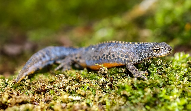
This fascinating time-lapse video from the Dutch director Jan van IJken tracks the development of a single-celled zygote into the hatched larva of an alpine newt. Captured in stunning detail at a microscopic level, BECOMING is a remarkable look at the process of cell division and differentiation, from where all animals—from newts to human beings—arise.
Becoming: the amazing journey from blob to salamander in 6 minutes
BECOMING is a short film about the miraculous genesis of animal life. In great microscopic detail, we see the ‘making of’ a salamander in its transparant egg from fertilisation to hatching.
The first stages of embryonic development are roughly the same for all animals, including humans. In the film, we can observe a universal process which normally is invisible: the very beginning of an animal’s life. A single cell is transformed into a complete, complex living organism with a beating heart and running bloodstream.
The alpine newt/salamander embryo (Ichthyosaura Alpestris) was followed very closely in a combination of timelapse and film. All stages of embryogenesis can be seen in this film: cleavage, gastrulation, neurulation and organogenesis. Time was condensed from about 3 weeks to 6 minutes.
Source: JanVanIjke.com

What exactly is happening in this film?
Jan Van IJken was clever to choose an amphibious alpine newt. "You can look straight into the egg," he told Live Science. "They are transparent, and you can see the whole process." So, he contacted a salamander breeder and picked up a few dozen fertilised eggs.
But there are only a few hours between fertilisation and the first cell division, so van IJken had to race home and, like a microsurgeon, unfurl and unglue each egg from the leaf where the salamander mother had carefully stuck it. "Sometimes, I was just in time," van IJken said.
Then, he placed the eggs in water-filled petri dishes and took thousands of photos using a camera attached to a microscope for the next four weeks.
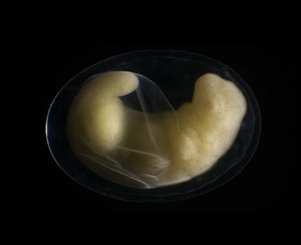
In that initial shot, you can see the fertilised egg (also called an embryo) in the clear, protective vitelline membrane. This membrane helps keep the egg moist and prevents pathogens from getting in.
In almost no time at all, the yellow salamander embryo has already divided into hundreds of cells.
Then, the embryo divides like crazy. Rather than expanding in size, the embryo increases its number of cells with every division, all within in that same amount of space.
There is a lag as each cell replicates the genetic material within it and then divides, in a process called mitosis.
At about the minute mark in the film, a "hole" appears in the embryo. The process that creates the hole is called gastrulation, when the embryo organises itself into three distinct cell layers.
At the gastrulation stage, the embryo consists of thousands of cells, and some already "know" that they, or their progeny, will become brain cells, gut cells or something else, but many of these cells are still on the outside of the egg.
During gastrulation, cells move around, organising themselves by going to the outermost layer, or ectoderm (nervous system, skin cells and pigment cells); the mesoderm (gut, muscles and red blood cells); or the inner layer, or endoderm (lung cells, thyroid cells and pancreatic cells).
Source: LiveScience.com
Continued below…
“It’s a true miracle, one cell dividing and then becoming this animal.” — Says filmmaker
At about the 1:50 mark, the embryo looks like it’s putting on a coat. This process is known as neurulation, and it happens as the neural tube rolls up. After this step, almost everything on the outside of the embryo is there to stay. This consists mostly of the organism’s protective skin.
At about 3 minutes into the video, you can see the limb buds forming. Soon enough, you can distinguish the head from the tail.
Van IJken stopped taking photos for the time lapse and switched to video as soon as the embryo moved, he noted.
Shortly after that, a tube that eventually becomes the heart begins to form—and once the heart beats, blood flows. You can even see the blood flowing through the gills, the structures that help the animal with gas exchange so that it can breathe underwater.
The developing salamander twitches as it gets older, likely because its growing brain is learning how to control the animal’s muscles.
Finally, the yellow tadpole breaks free from the protective membrane. It’s unclear how the animal knows when to do this, but hormones could play a role.
Watching the tadpoles hatch "was incredible" van IJken told Live Science. "How this internal clockwork makes the whole thing come to life, it’s incredible. It’s a true miracle, one cell dividing and then becoming this animal."
And the circle of life continues, he noted. After the tadpoles hatched, van IJken gave them back to the breeder and got to work editing the film — Check out van Ijken’s website for more stunning projects.
Source: LiveScience.com
